




AI-CVD: A New Paradigm for Comprehensive Cardiovascular and Metabolic Disease Risk Assessment Using Multidimensional AI-Enabled CT Biomarkers
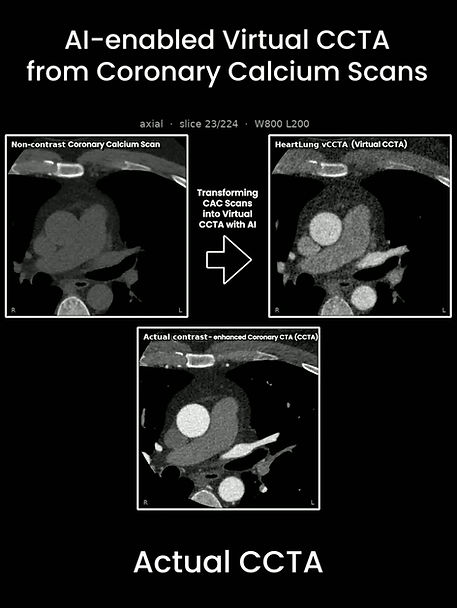
This review article presents AI-CVD as a multidimensional framework for extracting cardiovascular and cardiometabolic risk information from coronary artery calcium (CAC) scans and routine chest CT. Rather than relying on a single measurement, the platform brings together imaging biomarkers across the heart, vessels, lungs, liver, muscle, adipose tissue, and bone.
What the framework assesses
The manuscript describes AI-enabled assessment of coronary artery calcium and plaque features; cardiac chamber volumes and ratios; ventricular wall thickness and mass; aortic and mitral valve calcification; thoracic aortic and pulmonary artery dimensions; adiposity; liver attenuation; skeletal muscle volume and density; lung attenuation; and vertebral bone mineral density. These measurements are intended to provide a broader phenotype from imaging that may already be available in clinical care.
Potential clinical relevance
The authors summarize evidence from MESA, the Framingham Heart Study, and related cohorts linking selected AI-derived CT biomarkers with outcomes including coronary heart disease, heart failure, atrial fibrillation, stroke, diabetes, COPD, osteoporosis, and mortality. The review also describes integrated, endpoint-specific risk models; for example, it reports a 10-year AUC of 0.83 for AI-CVD-HF versus 0.79 for PREVENT-HF in cited work.
The proposed workflow uses imaging biomarkers to help identify patients whose scans show subclinical disease or adverse remodeling, while conventional risk factors and other actionable biomarkers help guide prevention and treatment. AI-CVD is positioned as a complement to established risk assessment, not a substitute for clinical evaluation.
Evidence status and next steps
The authors emphasize that much of the supporting evidence comes from observational cohorts. The manuscript calls for prospective validation, external testing across broader populations and imaging settings, and studies assessing whether AI-CVD-guided prevention changes clinical decisions and patient outcomes. As supplied, this is a submission manuscript marked “For Peer Review Only” (Submission ID 262522514), so its findings should be read as a review of an evolving evidence base rather than as a final peer-reviewed publication.
Article details
Nathan D. Wong, Kyle Atlas, Chenyu Zhang, Seyed Reza Mirjalili, Hamed Zarei, Soroush Masrouri, Oren Mechanic, Arthur Agatston, Robert A. Kloner, and Morteza Naghavi. “AI-CVD: A New Paradigm for Comprehensive Cardiovascular and Metabolic Disease Risk Assessment Using Multidimensional AI-Enabled CT Biomarkers.” Submitted to Future Cardiology. Submission ID: 262522514. Device Evaluation.


Comments