


AI-CVD Summary

AI-CVD Bundle
Coronary Calcification

_4_edited.jpg)
Aortic Calcification

Epicardial Fat
_5_edited.jpg)
Cardiac Chambers
_7_edited.jpg)
Aorta & PA Size
_10_edited.jpg)
Lung LAI
_9_edited.jpg)
Lung HAI
_11_edited.jpg)
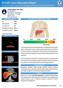
Liver Fat
_12_edited.jpg)
Muscle & Visceral Fat
_13_edited.jpg)
Bone Density


%20.png)


Explore AI-Powered Solutions & See Real AI Reports
See More. Detect Earlier. Treat Better. Earn More.

AI Biomarkers
Extract cardiovascular biomarkers from routine CT scans
Annotated Images
See AI segmentation and
overlays
Clinical Reports
Clear, actionable, and
physician-ready insights.
DICOM Preview
Interactive viewer and
comparison
Longitudinal Tracking
Monitor patient progress
over time
Risk Prediction
Estimate future cardiovascular disease and heart failure risk.


Coronary Arteric Calcification Findings


_5.jpeg)


_7.jpeg)


_9.jpeg)


_10.jpeg)


_11.jpeg)


%20.png)


_4.jpeg)
A Paradigm Shift in
CVD Risk Assessment
AI-CVD® is an advanced artificial intelligence technology that extracts quantitative imaging measurements from eligible cardiac and chest CT scans. This innovative approach significantly enhances CVD risk stratification by integrating imaging disease measures. AI-CVD® thus emerges as the most comprehensive tool that extends well beyond coronary artery calcium (CAC) score and coronary artery disease. AI-CVD® provides quantitative CT-derived measurements for clinician review. In research cohorts, several measurements have been associated with future cardiovascular outcomes.
Key Components of AI-CVD Developed by HeartLung.AI
Beyond the CAC Score
Aortic and pulmonary artery (PA) dimensions are important cardiovascular and pulmonary measurements. Changes in PA size can signal pulmonary hypertension or other vascular diseases. Accurate measurement through imaging helps in early detection and management. An enlarged aorta can indicate conditions like aneurysm or dissection. AI-CVD™ uses advanced algorithms to measure these sizes, enhancing risk assessment and timely intervention for cardiovascular and pulmonary conditions.


Myosteatosis, characterized by fat infiltration into skeletal muscle, is an emerging biomarker of systemic metabolic dysfunction. AI-driven measurement of thoracic skeletal muscle density from CAC scans has shown strong predictive value for HF, AF, CHD, and all-cause mortality. Recent studies demonstrate that combining myosteatosis with CAC scores amplifies risk prediction, particularly for males, making it a critical addition to AI-CVD®.


Thoracic vertebral bone density, measurable in routine CAC scans, provides an important marker for osteoporosis. Low BMD is independently associated with increased CVD risk and all-cause mortality, potentially mediated by shared inflammatory and metabolic pathways. Incorporating BMD into CVD risk assessments ensures early detection of both osteoporosis and its cardiovascular implications, enabling timely interventions.


Liver Fat
Fatty liver, detected opportunistically in CAC scans, correlates with metabolic syndrome, diabetes, and heightened CVD risk. By quantifying liver fat, AI-CVD™ identifies individuals at risk for cardiometabolic events, offering opportunities for early lifestyle or pharmacological interventions.


AVC, visible in CAC scans, predicts aortic stenosis and its associated cardiovascular outcomes. Early identification of AVC facilitates closer monitoring and timely surgical or medical interventions, reducing morbidity and mortality from valve-related complications.


Pulmonary emphysema, visible in chest CT scans, is linked to chronic obstructive pulmonary disease (COPD) and increased cardiovascular mortality. Including emphysema scoring in AI-CVD™ highlights the interplay between pulmonary and cardiovascular health, fostering integrated care approaches.


AI-driven analysis of cardiac chamber volumes—including the left atrium (LA), left ventricle (LV), right atrium (RA), and right ventricle (RV) provides actionable insights into heart failure and atrial fibrillation risk. Enlarged LA volume strongly predicts AF and stroke, while LV hypertrophy correlates with HF and coronary artery disease. These measurements add critical dimensions to CAC scans, improving predictive accuracy.


AI-CVD®: The Most Comprehensive CVD CT Screening
CVD Risk Assessment
AI-CVD® leverages advanced AI algorithms to enable physicians to evaluate cardiovascular disease risk that is otherwise missed. This approach significantly improves the prediction of conditions like heart failure and atrial fibrillation, outperforming traditional risk scores. Validated through studies like MESA and FHS, AI-CVD® aims to set a new standard in preventive cardiology.

