


_4_edited.jpg)

_5_edited.jpg)
_7_edited.jpg)
_10_edited.jpg)
_9_edited.jpg)
_11_edited.jpg)
_12_edited.jpg)
_13_edited.jpg)


%20.png)




AI-CAC: Coronary Plaque Analysis from Non-Contrast CT
Updated: Jul 17

AI-CAC introduces a novel artificial intelligence approach for coronary plaque analysis using non-contrast cardiac CT (CAC) scans, offering the potential to extract plaque characteristics traditionally obtainable only from contrast-enhanced coronary CT angiography (CCTA).
Conventional CCTA provides excellent visualization of coronary anatomy but requires intravenous contrast administration and is affected by scanner-, software-, and observer-related variability. AI-CAC addresses these limitations by applying advanced deep learning techniques to routine non-contrast CT scans, enabling detailed assessment of coronary plaque composition without the need for contrast agents.
The presented study validates AI-CAC against Cleerly AI-QCT using paired non-contrast CT and CCTA datasets from the Miami Heart Study. Results demonstrate strong agreement for multiple plaque measurements, including calcified plaque volume, total plaque volume, and lumen volume, highlighting the potential of AI-CAC as a practical and scalable alternative for coronary plaque assessment.
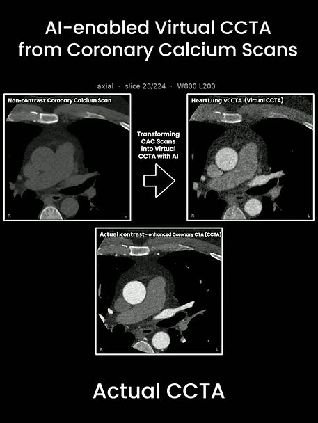
The presentation also introduces the next evolution of the platform—Virtual CCTA—an AI-driven technology capable of generating contrast-enhanced coronary images from standard non-contrast CT scans, opening new possibilities for accessible, lower-cost cardiovascular imaging and risk assessment.
Download the complete presentation below to explore the AI methodology, validation study, technical workflow, and the future vision of AI-CAC and Virtual CCTA.


Comments