


_4_edited.jpg)

_5_edited.jpg)
_7_edited.jpg)
_10_edited.jpg)
_9_edited.jpg)
_11_edited.jpg)
_12_edited.jpg)
_13_edited.jpg)


%20.png)




What cardiac insights are hiding in millions of routine chest CTs?
Updated: Aug 26

ESC 2026 · 10 Scientific Presentations · Oral & Poster · 6/10
CT vs. Echo: Unlocking More From Existing Imaging
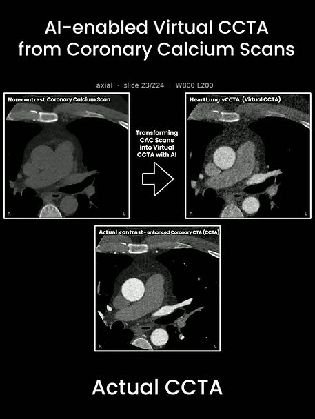
Approximately 19 million non-gated chest (lung) CT scans are performed in the U.S. each year for a wide range of clinical indications. These scans are not primarily acquired to evaluate cardiovascular disease - but could AI unlock cardiovascular information already captured within them?
At ESC Congress 2026, HeartLung.AI presented a study comparing AI-derived cardiac chamber volumetry from routine non-gated chest CT with echocardiography.
The study included 1,675 participants from the Multi-Ethnic Study of Atherosclerosis (MESA) who underwent both non-contrast, non-gated chest CT and echocardiography.
Researchers evaluated whether AI-derived cardiac chamber measurements from CT could provide meaningful information related to:
Heart failure
Atrial fibrillation
Cardiac chamber volumes
Long-term cardiovascular outcomes
The study directly compared the discrimination and prognostic value of CT-derived versus echocardiographic measurements for heart failure and atrial fibrillation.
Routine lung CT contains more than lung information.
With AI-enabled cardiac chamber volumetry, imaging already being performed for other clinical indications may offer an opportunity to extract additional cardiovascular insights - without requiring a dedicated cardiac CT acquisition.
📍 To be presented at ESC Congress 2026 · Munich
By Seyed Reza Mirjalili, MD
Download the full presentation below to explore the CT-versus-echocardiography comparisons, study results, and potential clinical implications.


Comments