


_4_edited.jpg)

_5_edited.jpg)
_7_edited.jpg)
_10_edited.jpg)
_9_edited.jpg)
_11_edited.jpg)
_12_edited.jpg)
_13_edited.jpg)


%20.png)




AI-enabled Cardiac Chambers Volumetry in Non-contrast Coronary Artery Calcium CT Scans vs. Contrast-Enhanced Coronary CT Angiography Scans in the Same Patients
The Multi-Ethnic Study of Atherosclerosis (MESA)
AUTHORS (FIRST NAME, LAST NAME):
Chenyu Zhang, Kyle Atlas, Song Shou Mao, Anthony Reeves, Matthew Budoff, Claudia Henschke, David Yankelevitz, and Morteza Naghavi.
Abstract:
Introduction:
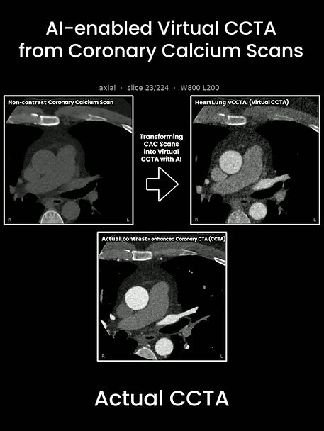
We have developed an AI-enabled automated volumetry of cardiac chambers (AutoChamber) that works both on non-contrast ECG-gated cardiac CT scans used for coronary artery calcium (CAC) score, and non-contrast non-gated lung cancer screening CT scans. We have recently reported the agreement between AutoChamber measurements vs cardiac MRI measurements. Here we report AutoChamber results on non-contrast CAC scans and contrast enhanced coronary CT angiography (CTA).
Methods:
We have obtained CT images from a previously published study in which 131 cases underwent non-contrast CAC and contrast enhanced CCTA at the Harbor UCLA Lundquist Research Institute. The cases consisted of 52.4% women with ages 55.0 ± 10.1 years. The cardiac chambers volume in 131 cases of contrast enhanced CCTA were measured using automated chamber volumetry by Philips Brilliance Volume v 4.5; Philips Healthcare.
Results:
The correlations between Philips’ automated chamber volumetry in contrast enhanced CCTA scans versus AutoChamber automated volumetry in non-contrast CAC scans for left atrium (LA), left ventricle (LV), right atrium (RA), right ventricle (RV), and left ventricular wall (LVW) volume were 0.73, 0.65, 0.85, 0.93, 0.90, respectively (P for all <0.01).
Conclusion:
AI-enabled AutoChamber volumetry based on non-contrast cardiac scans is feasible, and correlates reasonably well with contrast-enhanced cardiac CT scans. Further studies are warranted to reproduce these results and explore the add-on value of AutoChamber to CAC scans.


Comments